Fiber training: theory without practice?
Author:
Julio Valero
Published on:
1/23/2025

Do the anatomical and physiological properties of a muscle determine its adaptive response to different loading protocols?
There is a widespread belief that resistance training with different repetition ranges and loads induces differential activation of muscle fibers. A study published two years ago explored this hypothesis, providing empirical evidence on muscle plasticity in response to various training protocols.
Overview
Variables measured: The muscle thickness of the soleus and gastrocnemius was assessed by B-mode ultrasound, and the maximum isometric strength of the calf was assessed by dynamometry, using light loads (20-30 maximum repetitions) and heavy loads (6-10 maximum repetitions).
The results showed small variations between muscle groups and training protocols. The LIGHT protocol showed a slight advantage in terms of hypertrophy, while the HEAVY protocol showed a slight superiority in strength gains.
The results indicate that the way weight is distributed in exercises does not affect the growth of different types of muscle fibers in the calf. In addition, this muscle responds equally to different training routines, as long as they are performed at maximum intensity.
What's the problem?
Skeletal muscle cells, or muscle fibers, are grouped into bundles and form muscles. These fibers are primarily classified into type I and type II, differing in their speed of contraction, resistance to fatigue, and metabolism. Other properties such as size, strength, and mitochondrial density also vary between types. Type I muscle fibers are ideal for endurance, contracting slowly and resisting fatigue. Type II, on the other hand, are explosive but fatigue quickly, making them optimal for fast, powerful movements. Type II muscle fibers are more likely to grow in response to strength training, which explains why endurance athletes are typically less bulky. Although most muscles have a balanced mix of type I and II fibers, their proportion varies by muscle. For example, the triceps surae (calf) is a predominantly type II muscle, as shown in Figure 1.
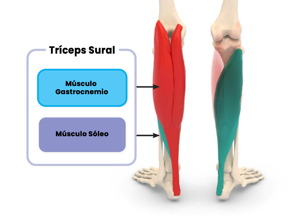
The gastrocnemius covers the soleus, both forming the triceps surae.
The gastrocnemius, composed of fast and slow fibers, forms the superficial part of the calf. Below, the soleus, predominantly slow fibers, completes this muscle. This fiber diversity makes the calf an ideal model to study how different types of training influence the growth of each fiber type, as demonstrated in this study.
Traditionally, it has been believed that the type of muscle fiber being worked depends on the load and repetitions used. However, recent research suggests that this relationship is not as clear as previously thought. Studies such as that of Grgic and Schoenfeld (2018) indicate that training to muscle failure, rather than the load itself, could be the determining factor in muscle hypertrophy. The results of this new study reinforce this idea, suggesting that differences in loading patterns do not have a significant impact on the specific muscle adaptations of each fiber type.
Purpose
The main objective was to analyze the differences in muscular adaptations (strength and hypertrophy) induced by two different repetition ranges (20-30 RM and 6-10 RM) during the execution of the calf raise exercise, using resistance training.
The authors also set out to analyze hypertrophy adaptations at the muscle fiber level in each of the calf muscles, in order to determine whether the proportion of fast- and slow-twitch fibers influences the magnitude and type of hypertrophic response.
Hypothesis
The researchers hypothesized that the high-intensity protocol would produce more significant neural and muscular adaptations, manifested in a greater increase in maximum strength and a preferential development of type II fibers in the gastrocnemius muscles. On the other hand, the low-intensity protocol would be associated with an increase in soleus muscle volume, due to hypertrophy of type I fibers.
What Did They Test and How?

Participants
At baseline, 30 healthy, untrained men participated in the study. A total of four subjects withdrew from the study for personal reasons (n = 2), noncompliance (n = 1), and injury (n = 1). Thus, 26 subjects completed the study.
Study procedures
This study employed a randomized intergroup experimental design. This means that each participant served as his or her own control, training each leg with a different protocol: LIGHT (20-30 repetitions maximum) and HEAVY (6-10 repetitions maximum). Leg assignment to each protocol was determined randomly for each individual.
The 8-week, supervised training program focused on calf raise exercises, both seated (leg flexed) and standing (leg extended), and was performed in two non-consecutive weekly sessions. To ensure adequate familiarization with the exercises, participants completed one week of training prior to the exercise with progressive loads and a variable number of repetitions.
During the training sessions, four sets per exercise were performed, with each set being performed to muscle failure. Rest times between sets and exercises were carefully controlled to optimize recovery and performance.
To minimize bias and ensure the objectivity of the results, two levels of blinding were implemented: the investigator performing the ultrasound measurements was unaware of the protocol assignment to each leg, and the statistical analysis was performed by an independent investigator who was also unaware of this assignment.
Table 1 Training protocol.
Session 1 | Load | Session 2 | Load | Session 3 | Load | Session 4 | Load |
Straight-leg calf raise | Light | Bent-leg calf raise | Heavy | Bent-leg calf raise | Light | Straight-leg calf raise | Heavy |
Straight-leg calf raise | Heavy | Bent-leg calf raise | Light | Bent-leg calf raise | Heavy | Straight-leg calf raise | Light |
Bent-leg calf raise | Light | Straight-leg calf raise | Heavy | Straight-leg calf raise | Light | Bent-leg calf raise | Heavy |
Bent-leg calf raise | Heavy | Straight-leg calf raise | Light | Straight-leg calf raise | Heavy | Bent-leg calf raise | Light |
The protocol was repeated four times for a total of 16 sessions
Straight-leg calf raise = standing calf raise; Bent-leg calf raise = seated calf raises.
Measures
Dietary adherence was assessed through self-reported food records in the MyFitnessPal app. These records were collected one week prior to the start of the training protocol (familiarization phase) and during the last week of the training protocol.
Anthropometric variables of weight and fat percentage were determined using an InBody 770 body composition analyzer.
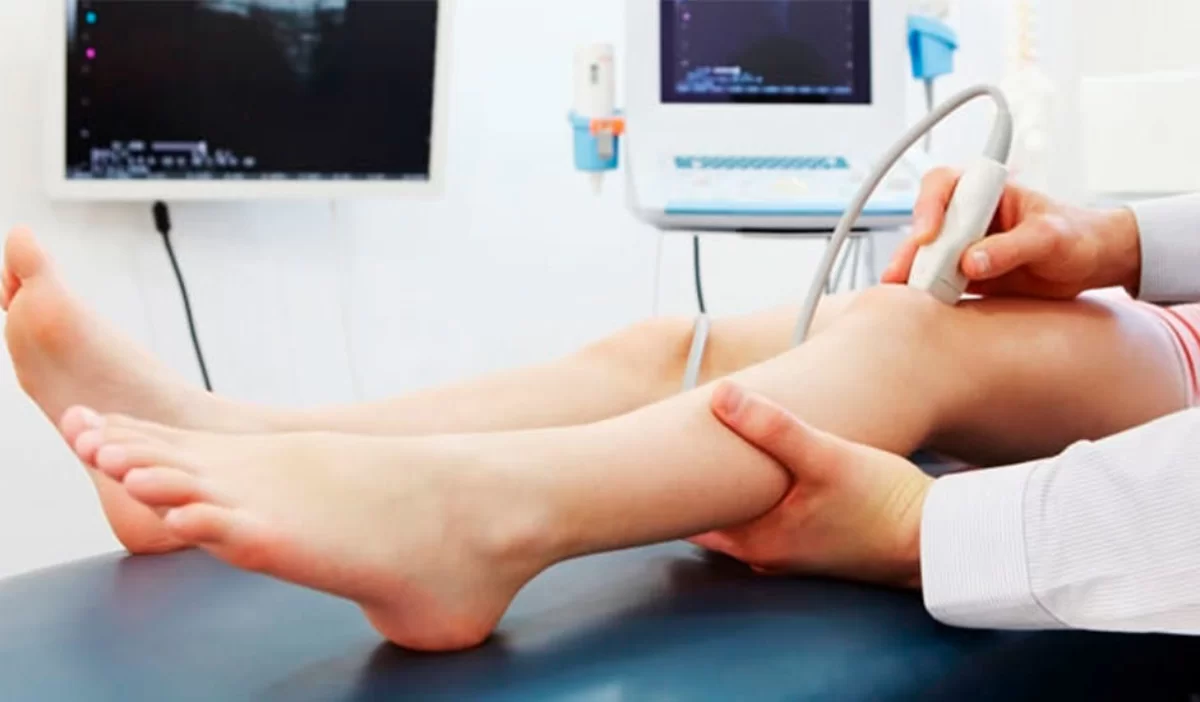
Muscle thickness (MT) of the soleus, medial gastrocnemius (MG), and lateral gastrocnemius (LG) muscles was quantified using B-mode ultrasound.
Maximal isometric calf strength was assessed using dynamometry. Each test consisted of a sustained contraction for 5 seconds, followed by a 30-second rest. Four repetitions of this protocol were performed to determine the maximum net joint moment.
¿What Did They Find?
Muscle Growth
No significant differences in muscle growth were observed between the muscles tested (soleus, medial and lateral gastrocnemius) or between training conditions (HEAVY vs. LIGHT). However, the lateral gastrocnemius showed the greatest increase in thickness, followed by the medial gastrocnemius and soleus. Detailed data are presented in Table 2.
Table 2 Effect of condition on within-muscle growth and isometric strength
Heavy Pre | Heavy Post | Heavy change | Light Pre | Light Post | Light Change | Between training condition effect size | |
Soleus (mm) | 18.8 ± 4.4 | 20.1 ± 4.6 | 1.3 ± 14 | 18.2 ± 4.3 | 19.7 ± 4.6 | 1.5 ± 1.3 | 0.2 (-0.3, 0.7) |
Medial gastrocnemius (mm) | 18.3 ± 3.2 | 19.7 ± 3.1 | 1.5 ± 1.3 | 17.7 ± 3.0 | 19.5 ± 3.0 | 1.8 ± 1.6 | 0.2 (-0.2, 0.8) |
Lateral gastrocnemius (mm) | 15.9 ± 2.6 | 17.9 ± 2.5 | 2.1 ± 1.4 | 15.6 ± 2.8 | 17.9 ± 3.2 | 2.3 ± 2.2 | 0.2 (-0.5, 0.8) |
Isometric plantar flexion (N-m) | 154 ± 48 | 170 ± 41 | 15 ± 37 | 153 ± 47 | 168 ± 41 | 15 ± 50 | -1.2 (-7.4, 4.5) |
Muscle strength
As with hypertrophy, changes in muscle strength were insignificant and showed no significant differences between groups. Table 2 details the results for isometric plantar flexion, where the HEAVY condition showed a slight increase of 1.2 N m.
What Do the Findings Mean?
If the theory of specific training for each type of fibre were correct, we would expect the soleus (mostly type I) to respond better to light training, while the gastrocnemius (mixed) to respond better to heavy training. However, our results show similar muscle growth in both muscles, regardless of the load. Although the gastrocnemius, with greater hypertrophic potential, showed slightly greater growth, the differences were minimal and favoured light training. This suggests that other factors, such as muscle functions and anatomical characteristics, could influence hypertrophy more than fibre type.
A limitation of the study is the indirect measurement of fiber hypertrophy using ultrasound rather than biopsies. Although the use of ultrasound is acceptable to compare muscles with different fiber composition, single-point measurements limit the generalizability of the results. Furthermore, isometric strength results might not fully reflect adaptations to heavier loads. Finally, the effect of cross-training might have influenced the observed strength adaptations.
Our results corroborate previous studies suggesting that muscle hypertrophy is similar regardless of the load used in training. Despite this, evidence on muscle fiber type-specific hypertrophy is limited and contradictory. While theoretically possible, practice shows that it is difficult to achieve in most cases. Further research is needed to clarify this point, but so far, there does not seem to be any significant advantage in training with fiber type-specific loads.
How Can You Apply These Findings?
To maximize calf growth, intense training is essential. Both high reps (20-30) and low reps (6-10) can be effective, provided proper loading is applied. If you've hit a plateau, vary your rep range. Also, combine seated and standing calf raises to work both major calf muscles.

References
Schoenfeld BJ, Vigotsky AD, Grgic J, Haun C, Contreras B, Delcastillo K, et al. Do the anatomical and physiological properties of a muscle determine its adaptive response to different loading protocols? Physiological reports, 8(9), e14427.
Talbot J, Maves L. Skeletal muscle fiber type: using insights from muscle developmental biology to dissect targets for susceptibility and resistance to muscle disease. Wiley interdisciplinary reviews. Developmental biology, 5(4), 518–534.
Fry, A. C. (2004). The role of resistance exercise intensity on muscle fibre adaptations. Sports medicine, 34(10), 663-679.
van Wessel, T., de Haan, A., van der Laarse, W. J., & Jaspers, R. T. (2010). The muscle fiber type-fiber size paradox: hypertrophy or oxidative metabolism? European journal of applied physiology, 110(4), 665–694.
Johnson, M. A., Polgar, J., Weightman, D., & Appleton, D. (1973). Data on the distribution of fibre types in thirty-six human muscles. An autopsy study. Journal of the neurological sciences, 18(1), 111–129.
Gollnick, P. D., Sjödin, B., Karlsson, J., Jansson, E., & Saltin, B. (1974). Human soleus muscle: a comparison of fiber composition and enzyme activities with other leg muscles. Pflugers Archiv : European journal of physiology, 348(3), 247–255.
Ogborn, D., & Schoenfeld, B. J. (2014). The role of fiber types in muscle hypertrophy: implications for loading strategies. Strength & Conditioning Journal, 36(2), 20-25.
Schuenke, M. D., Herman, J. R., Gliders, R. M., Hagerman, F. C., Hikida, R. S., Rana, S. R., Ragg, K. E., & Staron, R. S. (2012). Early-phase muscular adaptations in response to slow-speed versus traditional resistance-training regimens. European journal of applied physiology, 112(10), 3585–3595.
Grgic, J., & Schoenfeld, B. J. (2018). Are the Hypertrophic Adaptations to High and Low-Load Resistance Training Muscle Fiber Type Specific?. Frontiers in physiology, 9, 402.
Morton, R. W., Oikawa, S. Y., Wavell, C. G., Mazara, N., McGlory, C., Quadrilatero, J., Baechler, B. L., Baker, S. K., & Phillips, S. M. (2016). Neither load nor systemic hormones determine resistance training-mediated hypertrophy or strength gains in resistance-trained young men. Journal of applied physiology (Bethesda, Md. : 1985), 121(1), 129–138.
Lim, C., Kim, H. J., Morton, R. W., Harris, R., Phillips, S. M., Jeong, T. S., & Kim, C. K. (2019). Resistance Exercise-induced Changes in Muscle Phenotype Are Load Dependent. Medicine and science in sports and exercise, 51(12), 2578–2585.
Vandervoort, A. A., & McComas, A. J. (1983). A comparison of the contractile properties of the human gastrocnemius and soleus muscles. European journal of applied physiology and occupational physiology, 51(3), 435–440.
Duysens, J., van Wezel, B. M., Prokop, T., & Berger, W. (1996). Medial gastrocnemius is more activated than lateral gastrocnemius in sural nerve induced reflexes during human gait. Brain research, 727(1-2), 230–232.
Schoenfeld, B. J., Grgic, J., Ogborn, D., & Krieger, J. W. (2017). Strength and Hypertrophy Adaptations Between Low- vs. High-Load Resistance Training: A Systematic Review and Meta-analysis. Journal of strength and conditioning research, 31(12), 3508–3523.
Gimmon, Y., Riemer, R., Oddsson, L., y Melzer, I. (2011). El efecto de la fatiga del músculo flexor plantar en el control postural. Journal of electromyography and kinesiology: revista oficial de la Sociedad Internacional de Kinesiología Electrofisiológica, 21(6), 922–928.
Comparte en redes sociales
Recent posts

A bad night's sleep: a reason to stay up even longer?

Creatine Effectiveness: What Does Science Say About Its Benefits?

Does meal timing help you lose fat?
Is your triceps press building muscle or holding you back?

Nutrition tailored to you: based on your genetic profile.

Carbohydrates: the key to an explosive workout.