The Science of Muscle: Discover How Muscles Are Built
Author:
Julio Valero
Published on:
1/2/2025

The mechanisms of muscle hypertrophy and their application to resistance training.
Over a decade ago, a thorough analysis of the scientific literature laid the groundwork for our understanding of muscle growth. However, constant advances in research have revolutionized our perspective on this topic.
Overview
Muscle hypertrophy, a process still not fully understood, has generated intense scientific debate. Dr. Brad Schoenfeld proposed three key mechanisms: mechanical tension, muscle damage and metabolic stress. Although mechanical tension is the most widely supported, the role of the other two remains a subject of study and controversy.
What's the problem?
While losing weight may be faster, building muscle requires patience and a targeted approach. Hypertrophy, the process of thickening muscle fibers, is critical to muscle growth. Understanding this mechanism allows us to design more effective training and nutrition programs to achieve optimal results. Although hyperplasia has been studied, evidence in humans is limited, so hypertrophy remains the primary goal for those looking to increase muscle mass.
Strengthen your knowledge: Learn the fundamentals of muscle anatomy
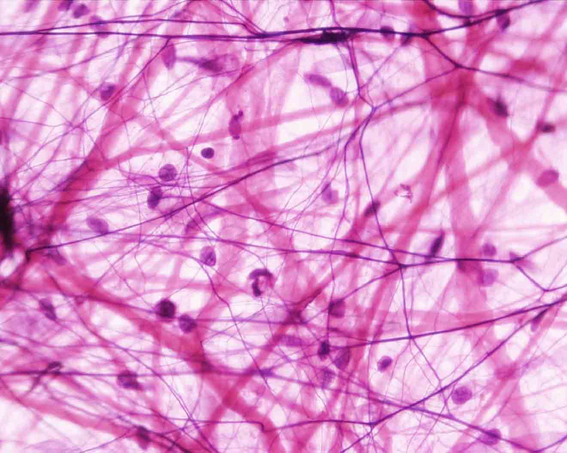
Imagine muscle tissue as a building. To understand how it grows and strengthens, we must first understand its bricks and how they are put together. Just as a building is made up of different materials and structures, muscle has levels of organization, from the smallest units to the entire muscle. Let’s start with the most basic level: the sarcomere, which is like a small brick that is repeated throughout the entire muscle structure. The sarcomere, the basic functional unit of the muscle, is like the tiny engine that powers movement. Composed primarily of actin and myosin filaments, these filaments slide past each other during muscle contraction, thereby shortening the sarcomere and, consequently, the entire muscle fiber.
The repetition of these sarcomeres along the myofibril creates the characteristic striated appearance of skeletal muscle. The myofibrils, in turn, are embedded in the sarcoplasm, the cytoplasm of the muscle fiber, and surrounded by the sarcolemma, its plasma membrane.
Several myofibrils group together to form a muscle fiber, which is a multinucleated cell. A bundle of muscle fibers, surrounded by a layer of connective tissue called the endomysium, forms a fascicle. The fascicles, in turn, group together to form the entire muscle, surrounded by an outermost layer of connective tissue called the epimysium.

Muscle fibers are like little factories of movement. Inside them, we find myofibrils, which are responsible for contracting. In addition, there are mitochondria (which produce energy), a sarcoplasmic reticulum (which stores calcium), nuclei (which control the cell) and a liquid called sarcoplasm. The interesting thing is that muscle fibers have many nuclei, unlike other cells that only have one.

Satellite Cells
Satellite cells, often referred to as resident muscle stem cells, are found in a quiescent state within the muscle fiber niche. Upon undergoing an intense and sustained mechanical stimulus, such as strength training, these cells are activated, proliferate, and fuse with existing muscle fibers. This process, known as myogenesis, leads to an increase in the number of nuclei within the muscle fiber.
The myonuclear dominance theory proposes that each muscle nucleus controls a volume of cytoplasm and its protein synthesis capacity. Therefore, an increase in the number of nuclei, thanks to the contribution of satellite cells, allows a greater potential for the synthesis of contractile proteins, such as actin and myosin, essential for muscle growth. This increase in protein synthesis leads to a thickening of the myofibrils and, consequently, to muscle hypertrophy.
Types of muscle hypertrophy
Myofibrillar
Imagine myofibrillar hypertrophy as the construction of a building. The bricks represent sarcomeres, the basic units of muscle structure. By adding more bricks (sarcomeres) in parallel, the building (the muscle fiber) becomes wider and stronger. This is similar to what happens when we lift weights and our muscle fibers become thicker. On the other hand, adding bricks in series would be like building more floors in the building, which would increase its height but not necessarily its width. However, in the context of muscle hypertrophy, growth in parallel is the predominant mechanism.
Sarcoplasmic
Sarcoplasmic hypertrophy involves not only an increase in cell size, but also changes in the biochemical composition of the muscle. A larger sarcoplasmic volume provides a larger physical space for the location of mitochondria, which may improve the oxidative capacity of the muscle and increase resistance to fatigue. Furthermore, the increase in glycogen content may provide an additional energy source for high-intensity exercise. It has been proposed that sarcoplasmic hypertrophy could play a crucial role in the early stages of muscle growth. By increasing cell volume, space is created for the addition of new myofibrils and the proliferation of satellite cells, the progenitor cells of the muscle, is facilitated. Once a certain threshold of sarcoplasmic growth has been reached, satellite cells can fuse with existing muscle fibers, donating new nuclei and allowing for increased synthesis of contractile proteins.
Connective tissue
Connective tissue hypertrophy is not an isolated phenomenon, but is closely linked to muscle remodeling processes. The extracellular matrix, in addition to providing structural support, acts as a microenvironment that influences muscle cell proliferation, differentiation, and function. Connective tissue cells, such as fibroblasts, interact with muscle fibers through cell adhesion molecules and growth factors, thereby regulating tissue homeostasis. During muscle hypertrophy, a complex interaction between muscle cells and connective tissue occurs. The increased mechanical demand on the muscle stimulates fibroblast proliferation and the synthesis of extracellular matrix proteins, resulting in a thickening of the connective tissue layers. This adaptive response seeks to strengthen the muscle structure and prevent injury.
Hormones
Hormones are chemical messengers that circulate in the bloodstream and influence various bodily functions. Anabolic hormones, such as testosterone, play a crucial role in muscle growth by promoting protein synthesis, the process of creating new muscle tissue.
Insulin-like growth factor 1
The pituitary gland, a small gland located in the brain, produces growth hormone (GH). This hormone is like a superhero for our muscles, as it helps repair them and grow after exercise. When we exercise intensely, our body produces a substance called lactate, which acts as a signal for the pituitary gland to release more GH. So training hard is like giving our growth hormone a boost!
Growth hormone (GH) acts as a potent modulator of metabolism, exerting both catabolic and anabolic effects. On the one hand, it stimulates lipolysis, i.e. the breakdown of fats, thus providing an alternative source of energy for the muscle. On the other hand, GH promotes protein synthesis, especially collagen, which contributes to the growth and repair of tissues, including muscle connective tissue. Although GH does not act directly on muscle contractile proteins, its effect on the extracellular matrix is essential for muscle growth and strength.
The benefits of GH for increasing muscle mass have been widely studied, especially in animal models. However, the results in humans are more complex and often contradictory. While GH undoubtedly stimulates the production of IGF-1, a potent promoter of muscle growth, its direct effect on protein synthesis in human muscles is still not completely clear. The lack of longitudinal studies in humans and individual differences in response to GH make it difficult to draw definitive conclusions.
The hormone hypothesis
The role of hormones in muscle hypertrophy has been the subject of intense debate in the scientific community. While it is widely accepted that hormones such as testosterone and insulin-like growth factor-1 (IGF-1) play a role in regulating muscle growth, their relative importance and the precise mechanisms by which they exert their effects remain a matter of research.
The hormonal hypothesis postulates that increases in circulating levels of these hormones in response to exercise are essential to stimulate muscle protein synthesis and, therefore, hypertrophy. However, this perspective has been challenged by studies suggesting that mechanical stress-induced activation of intracellular signaling pathways is the primary stimulus for muscle growth. These researchers argue that while hormones may potentiate the effects of training, they are not strictly necessary for hypertrophy.
The experimental evidence in this field is complex and often contradictory. Some studies have shown a positive correlation between hormone levels and muscle mass gains, while others have found no such association. Furthermore, the influence of individual factors, such as genetics, age and nutritional status, can moderate the hormonal response to exercise and consequently affect muscle growth.
Exercise-induced muscle hypertrophy
Schoenfeld proposed three key mechanisms for muscle hypertrophy: mechanical tension, muscle damage, and metabolic stress. Although these mechanisms have been widely studied, their relative contribution remains debated. It is important to remember that these models are simplifications of complex biological processes and do not accurately predict muscle growth in each individual. Imagine your muscle as a bank account. When you deposit more protein (synthesis) than you withdraw (breakdown), your account grows (hypertrophy). Stimuli such as weight training are like large deposits that make your muscle grow. If you withdraw more than you deposit, your muscle shrinks.

Mechanical stress
The extracellular matrix, a network of proteins and molecules surrounding muscle cells, plays a crucial role in transducing mechanical signals and regulating muscle growth. Forces applied to the muscle are not only transmitted through sarcomeres and costameres, but also interact with the extracellular matrix, generating a series of mechanical signals that influence muscle cell behavior.
The stiffness, viscosity, and composition of the extracellular matrix can modulate the response of muscle cells to mechanical stress. In addition, the extracellular matrix contains a variety of signaling molecules that can interact with cell surface receptors, amplifying mechanical signals and promoting muscle growth.
Imagine that mechanosensors are the alarm sensors of a building. When an alarm is triggered (muscle tension), a series of coordinated actions (myogenic pathways) are initiated to repair the damage (catabolism) and strengthen the structure of the building (anabolism). In the case of muscle, this repair and strengthening process leads to muscle growth.
Primary anabolic signaling pathways
The mechanical stress generated during weightlifting induces a series of intracellular events that culminate in the activation of anabolic signaling pathways. Mechanosensors located in the cell membrane and cytoskeleton detect the stress and transmit the signal to the interior of the cell. Through a complex network of molecular interactions, transcription factors are activated that promote the expression of genes related to muscle protein synthesis, such as growth factors and myokines.
PI3K/Akt y mTOR
The PI3K/Akt/mTOR pathway is a central signaling cascade in the regulation of cell growth and survival, including muscle hypertrophy. Activation of PI3K by various stimuli, such as growth factors, leads to phosphorylation and activation of Akt. In turn, Akt phosphorylates and activates mTOR, a master regulator of cell growth. mTOR promotes protein synthesis through activation of the mTORC1 pathway, which stimulates the formation of new ribosomes and mRNA translation, processes essential for muscle growth.
Mitogen-activated protein kinase
Mitogen-activated protein kinase (MAPK) is a key player in muscle growth. When we exercise, MAPK is activated and triggers a series of reactions that stimulate the production of new muscle proteins, which in turn increases the size of our muscles.
Calcium-regulated pathways
Calcineurin, a calcium-activated enzyme, influences muscle hypertrophy. Although research results are mixed, its role in regulating muscle mass is clear. In addition, other calcium-dependent enzymes may also be involved.
Muscle growth is like a secret message that is sent to our cells. When we lift weights, we send a signal to the muscle cells, telling them that they need to grow. This signal, like a letter, travels inside the cell and activates a series of processes that cause the cell to produce more proteins to become bigger. Although the details of this process are complex, the important thing to understand is that exercise is the message that starts it all.
Muscle damage
Schoenfeld subsequently questioned some of the central ideas about muscle damage, and new research has since continued to challenge this hypothesis.
What is muscle damage?
The work of Schoenfeld and other researchers has shown that muscle damage is a normal physiological process in response to strength training. Understanding the mechanisms underlying muscle damage is critical to designing more effective training programs and minimizing injury risk.
By performing eccentric exercises, such as negative reps, a greater degree of muscle damage can be induced and therefore stimulate a greater adaptive response. However, it is important to note that excessive muscle damage can delay recovery and increase the risk of injury. Therefore, it is crucial to find a balance between stimulus and stress to optimize training results.
The concept of the "repeated bout effect" suggests that gradual progression of training load and volume is critical to maximizing long-term results. By gradually increasing training intensity and volume, the muscle is allowed to adapt and become more resistant to damage.
In conclusion, muscle damage is a natural and necessary process for muscle growth. By understanding the mechanisms underlying muscle damage and applying the principles of strength training, we can design personalized and effective training programs to achieve our fitness goals.
While strength training can cause muscle damage, this damage isn't necessarily a bad thing. In fact, the process of muscle repair and regeneration after a training session is thought to be what stimulates muscle growth. However, the relationship between muscle damage and hypertrophy is complex and still not fully understood.

Human evidence analyzing muscle damage and hypertrophy
Despite what many believe, the relationship between muscle damage and muscle growth is not as clear-cut as it seems. Some studies show that muscle growth can occur without a lot of damage, while others suggest that too much damage could be counterproductive. Even techniques like BFR show that muscle can be built with less damage. In short, while muscle damage plays a role, it is not the only determining factor for muscle growth.
Metabolic stress
We’ve all experienced that “pumped” feeling in the gym, that muscular fullness that makes us feel stronger. But what’s behind that feeling? Metabolic stress is the scientific term that describes the physiological changes that occur in our muscles during strength training. The buildup of metabolites like lactate and the decrease in intracellular pH are signals our body sends to indicate that we’re working our muscles to the limit. Although lactate has traditionally been considered a waste product, there is growing evidence to suggest that this metabolite may play an important role in stimulating muscle growth.
Blood flow restriction (BFR) training is an innovative technique that takes advantage of metabolic stress to maximize muscle growth. By limiting blood flow, lactic acid and other metabolites build up in the muscle, creating an environment that favors hypertrophy. This training modality allows for similar results to heavier loads, but with a lower risk of injury. That's why more and more people are incorporating BFR into their training routines.
Cell swelling
Cell swelling, a phenomenon induced by intense exercise, plays a crucial role in muscle hypertrophy. This increase in cell volume, attributable to the accumulation of metabolites and water influx, triggers a cascade of molecular events that promote muscle growth. Cell swelling is postulated to activate anabolic signaling pathways, such as mTOR, thus stimulating protein synthesis and muscle hypertrophy. Furthermore, cell swelling could act as a stress signal, inducing the expression of genes related to muscle repair and growth. Cotamers, stretch-sensitive protein structures, could act as sensors of cell swelling, amplifying anabolic signals.
How can you apply this information?
We understand that all this information about cellular mechanisms can sound a little overwhelming. The important thing to remember is that while theory is interesting, practice is what really matters. To gain muscle, you need to lift weights with sufficient intensity and frequency. This means gradually increasing the weight you lift and making sure that each workout is challenging enough.
There is no magic formula for muscle growth, but there are many resources and trainers that can help you find the training program that is best for you.
Muscle hypertrophy is the result of proper stimulation. While the underlying physiological mechanisms are complex, the foundation of any effective training program is lifting weights regularly and progressively. Follow these 4 principles to optimize your results:
Train to the limit: Perform few repetitions with a lot of weight, occasionally reaching muscle failure.
Train a variety of rep ranges and intensities: reps from 4 to 25 with intensities ranging from 30 to 85% of 1RM.
Build volume: 10 sets per muscle group weekly is a good starting point for most.
Customize your workout: Choose exercises that strengthen your weaker muscles and allow you to move freely.
Want to maximize your muscle gains? Include at least two training sessions per muscle group in your weekly routine!
References
Schoenfeld B. J. (2010). The mechanisms of muscle hypertrophy and their application to resistance training. Journal of strength and conditioning research, 24(10), 2857–2872.
Haun, C. T., Vann, C. G., Roberts, B. M., Vigotsky, A. D., Schoenfeld, B. J., & Roberts, M. D. (2019). A Critical Evaluation of the Biological Construct Skeletal Muscle Hypertrophy: Size Matters but So Does the Measurement. Frontiers in physiology, 10, 247.
Roberts, M. D., Haun, C. T., Vann, C. G., Osburn, S. C., & Young, K. C. (2020). Sarcoplasmic hypertrophy in skeletal muscle: A scientific “unicorn” or resistance training adaptation?. Frontiers in Physiology, 11, 816.
Vierck, J., O'Reilly, B., Hossner, K., Antonio, J., Byrne, K., Bucci, L., & Dodson, M. (2000). Satellite cell regulation following myotrauma caused by resistance exercise. Cell biology international, 24(5), 263–272.
Toigo, M., & Boutellier, U. (2006). New fundamental resistance exercise determinants of molecular and cellular muscle adaptations. European journal of applied physiology, 97(6), 643–663.
Moss, F. P., & Leblond, C. P. (1971). Satellite cells as the source of nuclei in muscles of growing rats. The Anatomical record, 170(4), 421–435.
Allen, D. L., Roy, R. R., & Edgerton, V. R. (1999). Myonuclear domains in muscle adaptation and disease. Muscle & nerve, 22(10), 1350–1360.
Paul, A. C., & Rosenthal, N. (2002). Different modes of hypertrophy in skeletal muscle fibers. The Journal of cell biology, 156(4), 751–760.
Kelley G. (1996). Mechanical overload and skeletal muscle fiber hyperplasia: a meta-analysis. Journal of applied physiology (Bethesda, Md. : 1985), 81(4), 1584–1588.
Haun, C. T., Vann, C. G., Osburn, S. C., Mumford, P. W., Roberson, P. A., Romero, M. A., Fox, C. D., Johnson, C. A., Parry, H. A., Kavazis, A. N., Moon, J. R., Badisa, V., Mwashote, B. M., Ibeanusi, V., Young, K. C., & Roberts, M. D. (2019). Muscle fiber hypertrophy in response to 6 weeks of high-volume resistance training in trained young men is largely attributed to sarcoplasmic hypertrophy. PloS one, 14(6), e0215267.
Goldspink G. (2005). Mechanical signals, IGF-I gene splicing, and muscle adaptation. Physiology (Bethesda, Md.), 20, 232–238.
Hameed, M., Lange, K. H., Andersen, J. L., Schjerling, P., Kjaer, M., Harridge, S. D., & Goldspink, G. (2004). The effect of recombinant human growth hormone and resistance training on IGF-I mRNA expression in the muscles of elderly men. The Journal of physiology, 555(Pt 1), 231–240.
Yang, S. Y., & Goldspink, G. (2002). Different roles of the IGF-I Ec peptide (MGF) and mature IGF-I in myoblast proliferation and differentiation. FEBS letters, 522(1-3), 156–160.
Wackerhage, H., Schoenfeld, B. J., Hamilton, D. L., Lehti, M., & Hulmi, J. J. (2019). Stimuli and sensors that initiate skeletal muscle hypertrophy following resistance exercise. Journal of applied physiology (Bethesda, Md. : 1985), 126(1), 30–43.
Harridge S. D. (2007). Plasticity of human skeletal muscle: gene expression to in vivo function. Experimental physiology, 92(5), 783–797.
Gobinet, J., Poujol, N., & Sultan, C. h. (2002). Molecular action of androgens. Molecular and cellular endocrinology, 198(1-2), 15–24.
Urban, R. J., Bodenburg, Y. H., Gilkison, C., Foxworth, J., Coggan, A. R., Wolfe, R. R., & Ferrando, A. (1995). Testosterone administration to elderly men increases skeletal muscle strength and protein synthesis. The American journal of physiology, 269(5 Pt 1), E820–E826.
Zhao, W., Pan, J., Zhao, Z., Wu, Y., Bauman, W. A., & Cardozo, C. P. (2008). Testosterone protects against dexamethasone-induced muscle atrophy, protein degradation and MAFbx upregulation. The Journal of steroid biochemistry and molecular biology, 110(1-2), 125–129.
Veldhuis, J. D., Keenan, D. M., Mielke, K., Miles, J. M., & Bowers, C. Y. (2005). Testosterone supplementation in healthy older men drives GH and IGF-I secretion without potentiating peptidyl secretagogue efficacy. European journal of endocrinology, 153(4), 577–586.
Kjaer M. (2004). Role of extracellular matrix in adaptation of tendon and skeletal muscle to mechanical loading. Physiological reviews, 84(2), 649–698.
Vingren, J. L., Kraemer, W. J., Ratamess, N. A., Anderson, J. M., Volek, J. S., & Maresh, C. M. (2010). Testosterone physiology in resistance exercise and training: the up-stream regulatory elements. Sports medicine (Auckland, N.Z.), 40(12), 1037–1053.
Spiering, B. A., Kraemer, W. J., Vingren, J. L., Ratamess, N. A., Anderson, J. M., Armstrong, L. E., Nindl, B. C., Volek, J. S., Häkkinen, K., & Maresh, C. M. (2009). Elevated endogenous testosterone concentrations potentiate muscle androgen receptor responses to resistance exercise. The Journal of steroid biochemistry and molecular biology, 114(3-5), 195–199.
Harridge S. D. (2007). Plasticity of human skeletal muscle: gene expression to in vivo function. Experimental physiology, 92(5), 783–797.
Sinha-Hikim, I., Cornford, M., Gaytan, H., Lee, M. L., & Bhasin, S. (2006). Effects of testosterone supplementation on skeletal muscle fiber hypertrophy and satellite cells in community-dwelling older men. The Journal of clinical endocrinology and metabolism, 91(8), 3024–3033.
Gharahdaghi, N., Phillips, B. E., Szewczyk, N. J., Smith, K., Wilkinson, D. J., & Atherton, P. J. (2021). Links Between Testosterone, Oestrogen, and the Growth Hormone/Insulin-Like Growth Factor Axis and Resistance Exercise Muscle Adaptations. Frontiers in physiology, 11, 621226.
Giustina, A., Mazziotti, G., & Canalis, E. (2008). Growth hormone, insulin-like growth factors, and the skeleton. Endocrine reviews, 29(5), 535–559.
Hartman, M. L., Iranmanesh, A., Thorner, M. O., & Veldhuis, J. D. (1993). Evaluation of pulsatile patterns of growth hormone release in humans: A brief review. American journal of human biology : the official journal of the Human Biology Council, 5(6), 603–614.
Kraemer, W. J., Marchitelli, L., Gordon, S. E., Harman, E., Dziados, J. E., Mello, R., Frykman, P., McCurry, D., & Fleck, S. J. (1990). Hormonal and growth factor responses to heavy resistance exercise protocols. Journal of applied physiology (Bethesda, Md. : 1985), 69(4), 1442–1450.
Kraemer, W. J., Fleck, S. J., Dziados, J. E., Harman, E. A., Marchitelli, L. J., Gordon, S. E., Mello, R., Frykman, P. N., Koziris, L. P., & Triplett, N. T. (1993). Changes in hormonal concentrations after different heavy-resistance exercise protocols in women. Journal of applied physiology (Bethesda, Md. : 1985), 75(2), 594–604.
Kragstrup, T. W., Kjaer, M., & Mackey, A. L. (2011). Structural, biochemical, cellular, and functional changes in skeletal muscle extracellular matrix with aging. Scandinavian journal of medicine & science in sports, 21(6), 749–757.
Doessing, S., Heinemeier, K. M., Holm, L., Mackey, A. L., Schjerling, P., Rennie, M., Smith, K., Reitelseder, S., Kappelgaard, A. M., Rasmussen, M. H., Flyvbjerg, A., & Kjaer, M. (2010). Growth hormone stimulates the collagen synthesis in human tendon and skeletal muscle without affecting myofibrillar protein synthesis. The Journal of physiology, 588(Pt 2), 341–351.
Sandri, M., Barberi, L., Bijlsma, A. Y., Blaauw, B., Dyar, K. A., Milan, G., Mammucari, C., Meskers, C. G., Pallafacchina, G., Paoli, A., Pion, D., Roceri, M., Romanello, V., Serrano, A. L., Toniolo, L., Larsson, L., Maier, A. B., Muñoz-Cánoves, P., Musarò, A., Pende, M., … Schiaffino, S. (2013). Signalling pathways regulating muscle mass in ageing skeletal muscle: the role of the IGF1-Akt-mTOR-FoxO pathway. Biogerontology, 14(3), 303–323.
Schiaffino, S., Dyar, K. A., Ciciliot, S., Blaauw, B., & Sandri, M. (2013). Mechanisms regulating skeletal muscle growth and atrophy. The FEBS journal, 280(17), 4294–4314.
Velloso C. P. (2008). Regulation of muscle mass by growth hormone and IGF-I. British journal of pharmacology, 154(3), 557–568.
Consitt, L. A., Saneda, A., Saxena, G., List, E. O., & Kopchick, J. J. (2017). Mice overexpressing growth hormone exhibit increased skeletal muscle myostatin and MuRF1 with attenuation of muscle mass. Skeletal muscle, 7(1), 17.
Sotiropoulos, A., Ohanna, M., Kedzia, C., Menon, R. K., Kopchick, J. J., Kelly, P. A., & Pende, M. (2006). Growth hormone promotes skeletal muscle cell fusion independent of insulin-like growth factor 1 up-regulation. Proceedings of the National Academy of Sciences of the United States of America, 103(19), 7315–7320.
Kraemer, W. J., Ratamess, N. A., & Nindl, B. C. (2017). Recovery responses of testosterone, growth hormone, and IGF-1 after resistance exercise. Journal of applied physiology (Bethesda, Md. : 1985), 122(3), 549–558.
Mangine, G. T., Hoffman, J. R., Gonzalez, A. M., Townsend, J. R., Wells, A. J., Jajtner, A. R., Beyer, K. S., Boone, C. H., Wang, R., Miramonti, A. A., LaMonica, M. B., Fukuda, D. H., Witta, E. L., Ratamess, N. A., & Stout, J. R. (2017). Exercise-Induced Hormone Elevations Are Related to Muscle Growth. Journal of strength and conditioning research, 31(1), 45–53.
Schoenfeld B. J. (2013). Postexercise hypertrophic adaptations: a reexamination of the hormone hypothesis and its applicability to resistance training program design. Journal of strength and conditioning research, 27(6), 1720–1730.
West, D. W., Burd, N. A., Staples, A. W., & Phillips, S. M. (2010). Human exercise-mediated skeletal muscle hypertrophy is an intrinsic process. The international journal of biochemistry & cell biology, 42(9), 1371–1375.
West, D. W., Kujbida, G. W., Moore, D. R., Atherton, P., Burd, N. A., Padzik, J. P., De Lisio, M., Tang, J. E., Parise, G., Rennie, M. J., Baker, S. K., & Phillips, S. M. (2009). Resistance exercise-induced increases in putative anabolic hormones do not enhance muscle protein synthesis or intracellular signalling in young men. The Journal of physiology, 587(Pt 21), 5239–5247.
Kvorning, T., Andersen, M., Brixen, K., & Madsen, K. (2006). Suppression of endogenous testosterone production attenuates the response to strength training: a randomized, placebo-controlled, and blinded intervention study. American journal of physiology. Endocrinology and metabolism, 291(6), E1325–E1332.
Morton, R. W., Sato, K., Gallaugher, M., Oikawa, S. Y., McNicholas, P. D., Fujita, S., & Phillips, S. M. (2018). Muscle Androgen Receptor Content but Not Systemic Hormones Is Associated With Resistance Training-Induced Skeletal Muscle Hypertrophy in Healthy, Young Men. Frontiers in physiology, 9, 1373.
Vierck, J., O'Reilly, B., Hossner, K., Antonio, J., Byrne, K., Bucci, L., & Dodson, M. (2000). Satellite cell regulation following myotrauma caused by resistance exercise. Cell biology international, 24(5), 263–272.
Bodine, S. C., Stitt, T. N., Gonzalez, M., Kline, W. O., Stover, G. L., Bauerlein, R., Zlotchenko, E., Scrimgeour, A., Lawrence, J. C., Glass, D. J., & Yancopoulos, G. D. (2001). Akt/mTOR pathway is a crucial regulator of skeletal muscle hypertrophy and can prevent muscle atrophy in vivo. Nature cell biology, 3(11), 1014–1019.
Blaauw, B., Schiaffino, S., & Reggiani, C. (2013). Mechanisms modulating skeletal muscle phenotype. Comprehensive Physiology, 3(4), 1645–1687.
Kramer, H. F., & Goodyear, L. J. (2007). Exercise, MAPK, and NF-kappaB signaling in skeletal muscle. Journal of applied physiology (Bethesda, Md. : 1985), 103(1), 388–395.
Crabtree G. R. (1999). Generic signals and specific outcomes: signaling through Ca2+, calcineurin, and NF-AT. Cell, 96(5), 611–614.
Hudson, M. B., & Price, S. R. (2013). Calcineurin: a poorly understood regulator of muscle mass. The international journal of biochemistry & cell biology, 45(10), 2173–2178.
Chin E. R. (2005). Role of Ca2+/calmodulin-dependent kinases in skeletal muscle plasticity. Journal of applied physiology (Bethesda, Md. : 1985), 99(2), 414–423.
Schoenfeld B. J. (2012). Does exercise-induced muscle damage play a role in skeletal muscle hypertrophy?. Journal of strength and conditioning research, 26(5), 1441–1453.
Damas, F., Libardi, C. A., & Ugrinowitsch, C. (2018). The development of skeletal muscle hypertrophy through resistance training: the role of muscle damage and muscle protein synthesis. European journal of applied physiology, 118(3), 485–500.
Malm C. (2001). Exercise-induced muscle damage and inflammation: fact or fiction?. Acta physiologica Scandinavica, 171(3), 233–239.
Kuipers H. (1994). Exercise-induced muscle damage. International journal of sports medicine, 15(3), 132–135.
Schott, J., McCully, K., & Rutherford, O. M. (1995). The role of metabolites in strength training. II. Short versus long isometric contractions. European journal of applied physiology and occupational physiology, 71(4), 337–341.
McHugh M. P. (2003). Recent advances in the understanding of the repeated bout effect: the protective effect against muscle damage from a single bout of eccentric exercise. Scandinavian journal of medicine & science in sports, 13(2), 88–97.
Schwane, J. A., Johnson, S. R., Vandenakker, C. B., & Armstrong, R. B. (1983). Delayed-onset muscular soreness and plasma CPK and LDH activities after downhill running. Medicine and science in sports and exercise, 15(1), 51–56.
Evans, W. J., & Cannon, J. G. (1991). The metabolic effects of exercise-induced muscle damage. Exercise and sport sciences reviews, 19, 99–125.
McKay, B. R., O'Reilly, C. E., Phillips, S. M., Tarnopolsky, M. A., & Parise, G. (2008). Co-expression of IGF-1 family members with myogenic regulatory factors following acute damaging muscle-lengthening contractions in humans. The Journal of physiology, 586(22), 5549–5560.
Howell, J. N., Chleboun, G., & Conatser, R. (1993). Muscle stiffness, strength loss, swelling and soreness following exercise-induced injury in humans. The Journal of physiology, 464, 183–196.
Flann, K. L., LaStayo, P. C., McClain, D. A., Hazel, M., & Lindstedt, S. L. (2011). Muscle damage and muscle remodeling: no pain, no gain?. The Journal of experimental biology, 214(Pt 4), 674–679.
Damas, F., Phillips, S. M., Libardi, C. A., Vechin, F. C., Lixandrão, M. E., Jannig, P. R., Costa, L. A., Bacurau, A. V., Snijders, T., Parise, G., Tricoli, V., Roschel, H., & Ugrinowitsch, C. (2016). Resistance training-induced changes in integrated myofibrillar protein synthesis are related to hypertrophy only after attenuation of muscle damage. The Journal of physiology, 594(18), 5209–5222.
Loenneke, J. P., Thiebaud, R. S., & Abe, T. (2014). Does blood flow restriction result in skeletal muscle damage? A critical review of available evidence. Scandinavian journal of medicine & science in sports, 24(6), e415–e422.
Smith, R. C., & Rutherford, O. M. (1995). The role of metabolites in strength training. I. A comparison of eccentric and concentric contractions. European journal of applied physiology and occupational physiology, 71(4), 332–336.
Wells, G. D., Selvadurai, H., & Tein, I. (2009). Bioenergetic provision of energy for muscular activity. Paediatric respiratory reviews, 10(3), 83–90.
Lambert, C. P., & Flynn, M. G. (2002). Fatigue during high-intensity intermittent exercise: application to bodybuilding. Sports medicine (Auckland, N.Z.), 32(8), 511–522.
Jessee, M. B., Mattocks, K. T., Buckner, S. L., Dankel, S. J., Mouser, J. G., Abe, T., & Loenneke, J. P. (2018). Mechanisms of blood flow restriction: the new testament. Techniques in Orthopaedics, 33(2), 72-79.
Lang, F., Busch, G. L., Ritter, M., Völkl, H., Waldegger, S., Gulbins, E., & Häussinger, D. (1998). [Functional significance of cell volume regulatory mechanisms.](https://doi.org/10.1152/physrev.1998.78.1.247.) Physiological reviews, 78(1), 247–306.
Sjøgaard G. (1986). Water and electrolyte fluxes during exercise and their relation to muscle fatigue. Acta physiologica Scandinavica. Supplementum, 556, 129–136.
Sjøgaard, G., Adams, R. P., & Saltin, B. (1985). Water and ion shifts in skeletal muscle of humans with intense dynamic knee extension. The American journal of physiology, 248(2 Pt 2), R190–R196.
Low, S. Y., Rennie, M. J., & Taylor, P. M. (1997). Signaling elements involved in amino acid transport responses to altered muscle cell volume. FASEB journal : official publication of the Federation of American Societies for Experimental Biology, 11(13), 1111–1117.
Dangott, B., Schultz, E., y Mozdziak, PE (2000). La suplementación dietética con monohidrato de creatina aumenta la actividad mitótica de las células satélite durante la hipertrofia compensatoria. Revista internacional de medicina deportiva , 21 (1), 13–16.
Schoenfeld, B., Fisher, J., Grgic, J., Haun, C., Helms, E., Phillips, S., ... y Vigotsky, A. (2021). Recomendaciones de entrenamiento de resistencia para maximizar la hipertrofia muscular en una población atlética: Posición de referencia de la IUSCA . Revista internacional de fuerza y acondicionamiento , 1 (1).
Comparte en redes sociales
Recent posts

A bad night's sleep: a reason to stay up even longer?

Creatine Effectiveness: What Does Science Say About Its Benefits?

Does meal timing help you lose fat?
Is your triceps press building muscle or holding you back?

Nutrition tailored to you: based on your genetic profile.

Carbohydrates: the key to an explosive workout.